- Written by Jakub Bartnik
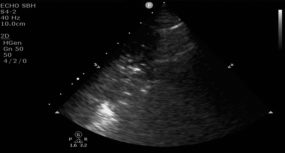
EMS calls ahead for a 35 year old male found altered and hypoxic in his apartment. Upon arrival, you note he is febrile, hypotensive, hypoxic on 100% oxygen to a spO2 of 70%. You decide to intubate after initiating fluid resuscitation, pressor support, and broad spectrum antibiotics. Your differential is broad and includes pulmonary embolism, pneumothorax, sepsis/pneumonia. After confirming ETT placement, you attempt to narrow your differential and place the phased array probe on the right anterior lung and find:

What do you see?
Here is another clip:
What’s your diagnosis?
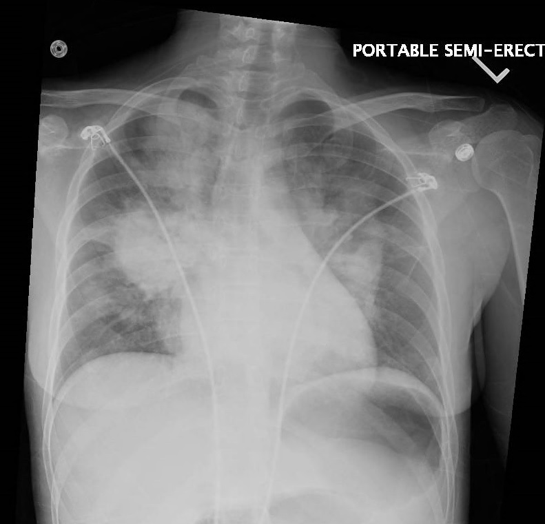
Here is the corresponding chest xray:
This is what pneumonia looks like on ultrasound.
Note the dynamic air bronchograms within the consolidated lung

There are two type of air bronchograms – static and dynamic.
Static bronchograms are nonspecific and may be seen in atelectasis.
Dynamic bronchograms are specific for pneumonia.
Check out this great 5minutesono from Jacob Avila on air bronchograms with some great examples.


